
MedGEN-Bench Review-Only Data Sample
This review-only dataset package contains 45 manually selected, unmodified benchmark records: 3 records for each of its 15 named tasks. It supports inspection of image pairs, task instructions, reference answers, construction-prompt evidence, and curation criteria. It is not a clinical dataset release and must not be redistributed.
Contents
records.jsonl— anonymized task records and image-file mappings.images/— native-resolution input/reference images for visual inspection.prompts/— exact record-level instructions plus documented construction/evaluation prompt material.rubric/— curation criteria and the review-attachment selection checklist.MANIFEST.json— coverage and SHA-256 integrity information.
The package intentionally contains no source-code or executable files.
Coverage
The package covers all 15 named tasks and the six canonical reporting modalities (CT, MRI, ultrasound, X-ray, pathology, and clinical photography). The counterfactual removal/reconstruction records are synthetic benchmark tasks; they are not clinical forecasting, diagnostic, or preprocessing recommendations.
Visual sample gallery
Open this file in a Markdown previewer. Each record retains its native image bytes in images/; the short answer below is a concise reviewer synopsis, while the canonical record answer is in records.jsonl.
VQA
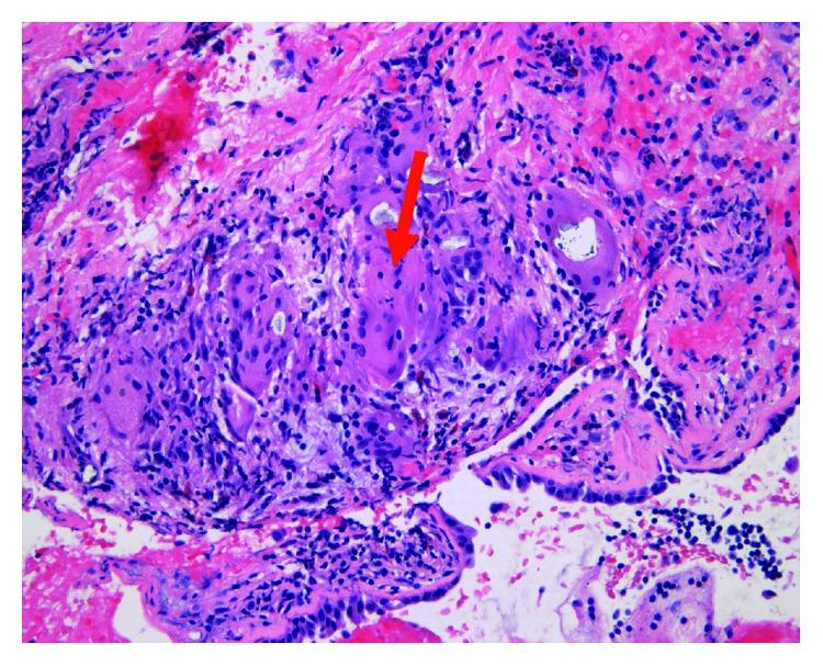
S01 — multiple-choice · Pathology
 —
—
Task. Question: What is the type of staining used in this image? Options: A. H&E B. PAS C. Trichrome D. Immunohistochemical
Expected output. H&E stain.
Selection note: Histopathology multiple-choice item: identify H&E staining.
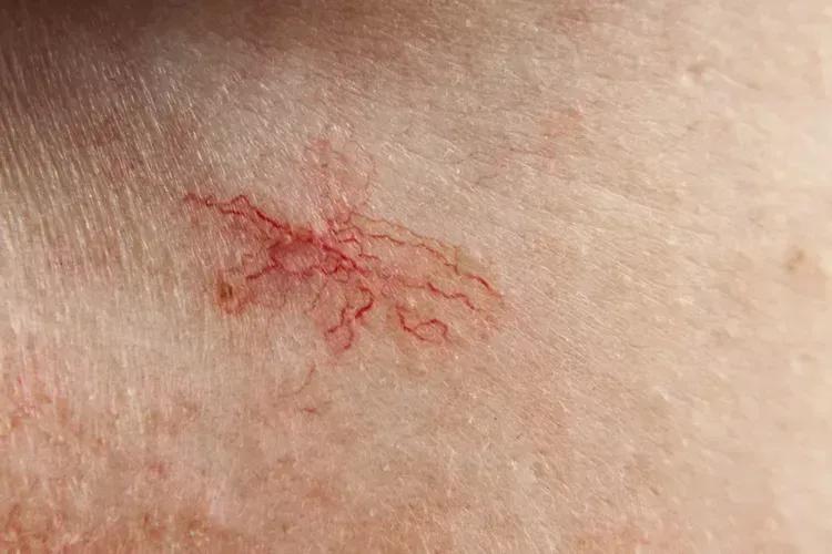
S02 — blank-filling · Clinical Photography
 —
—
Task. The skin condition shown is __.
Expected output. Spider angioma.
Selection note: Dermatology blank-filling item with a localized skin finding.
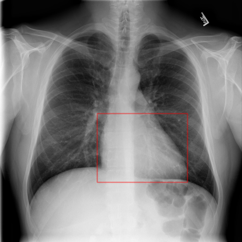
S03 — question-answering · X-ray
 —
—
Task. What organ is inside the red box in this image?
Expected output. The heart.
Selection note: Region-grounded radiograph question using a visible red box.
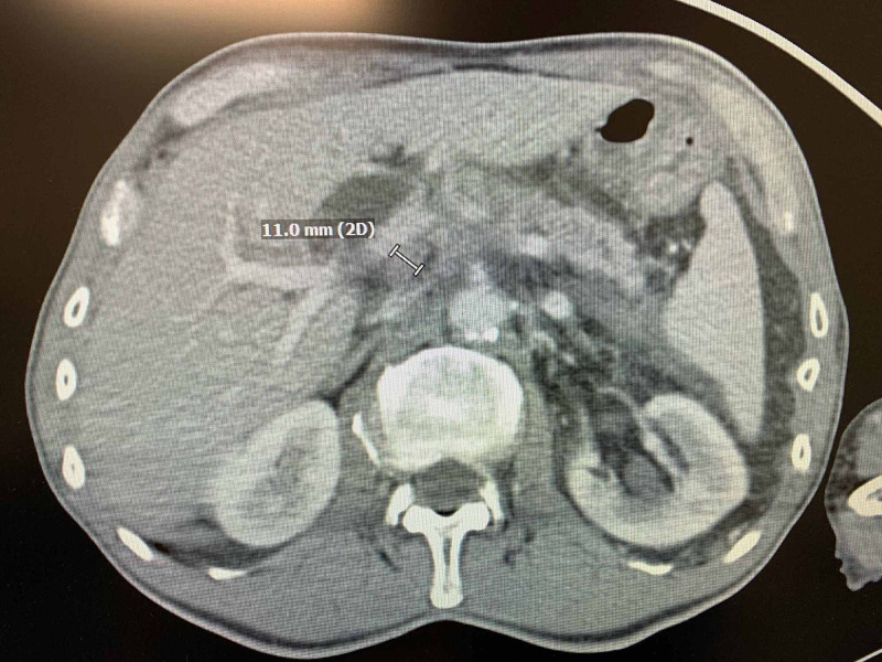
S04 — report-generation · CT
 —
—
Task. Generate a clinical report for this CT scan.
Expected output. Pancreatic head-neck mass with ductal dilatation and vascular involvement.
Selection note: CT report-generation example with a focal pancreatic finding.
ImageEditing
S05 — anatomical-annotation · Ultrasound
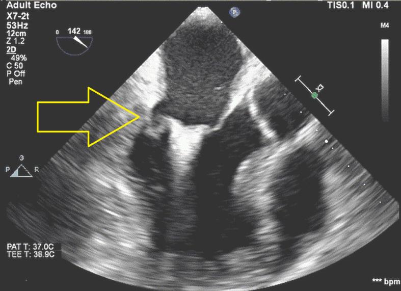
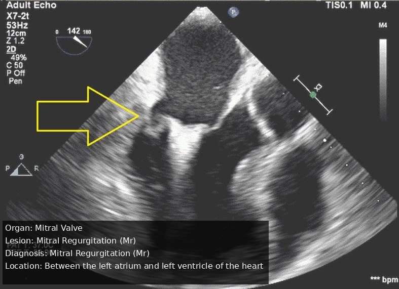
Task. Annotate this Ultrasound image of Mitral Valve with detailed labels for the Mitral Regurgitation (Mr), including anatomical landmarks and diagnostic findings
Expected output. Annotated mitral-valve ultrasound with diagnostic labels.
Selection note: Ultrasound annotation pair for mitral regurgitation.
S06 — artifact-removal · X-ray
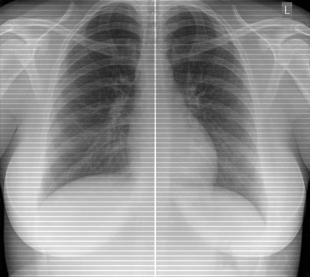
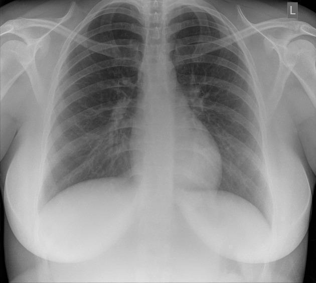
Task. Correct imaging artifacts in this X-Ray image
Expected output. PA chest radiograph with grid-line artifacts removed.
Selection note: Chest radiograph with visible grid-line artifacts and an artifact-reduced reference.
S07 — contrast-enhancement · Clinical Photography
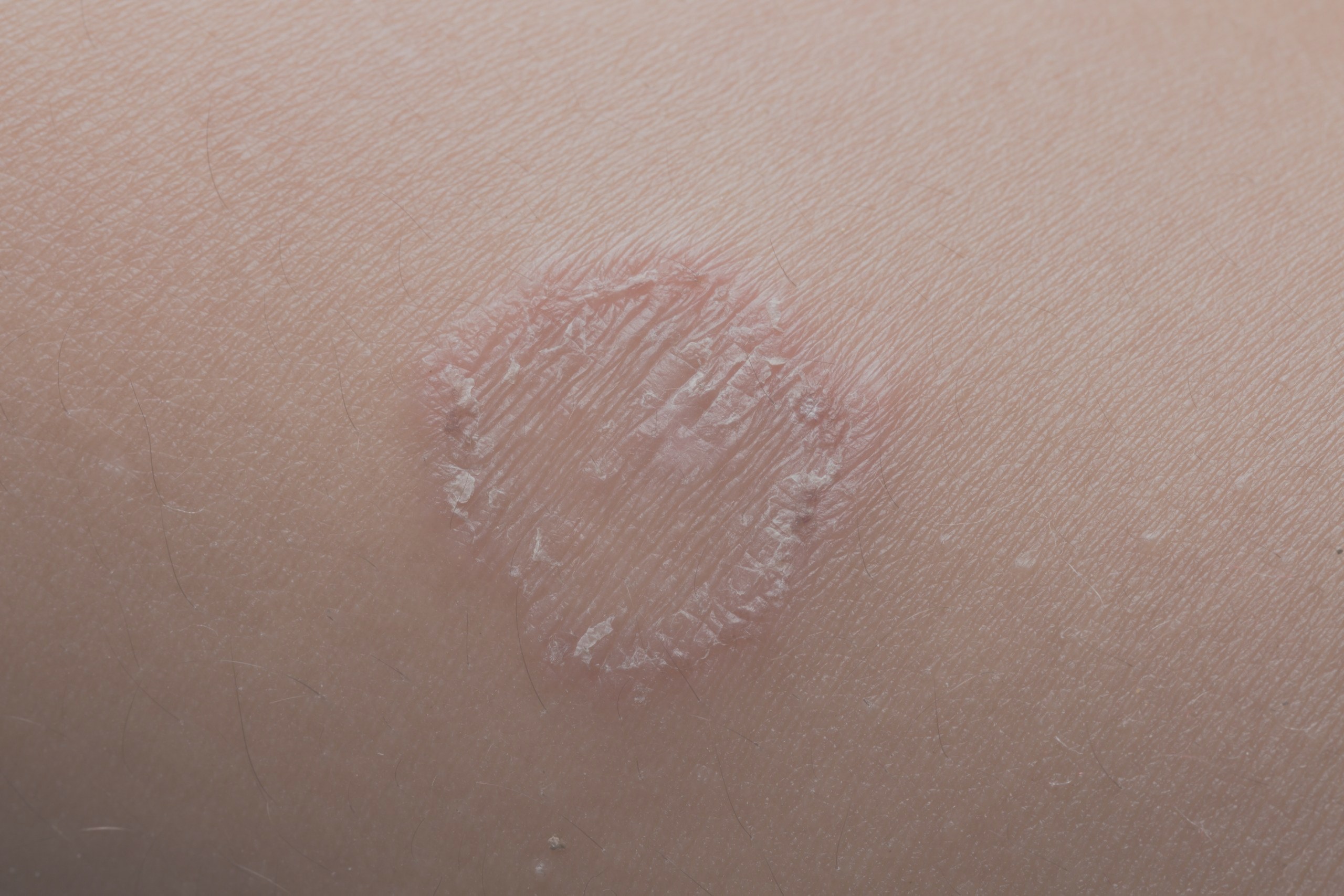
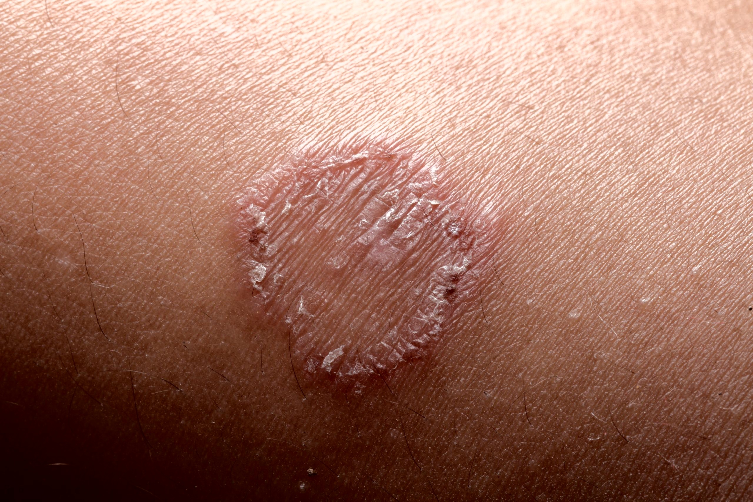
Task. Enhance the contrast of this Clinical Photo image using advanced image processing algorithms for improved diagnostic visibility
Expected output. Skin lesion with improved contrast and brightness.
Selection note: Dermatology contrast-enhancement pair; the image is non-identifying.
S08 — noise-reconstruction · CT
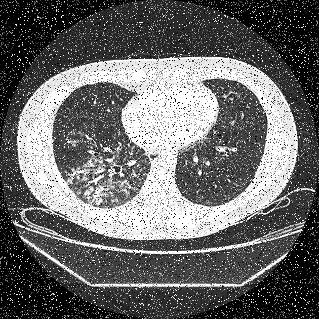
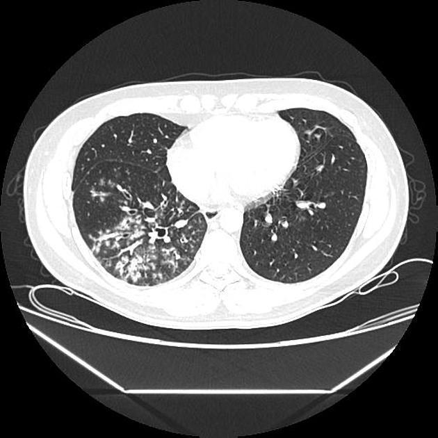
Task. Apply sophisticated noise reduction to this CT scan while preserving fine anatomical details and diagnostic features
Expected output. Denoised chest CT with preserved pulmonary and mediastinal detail.
Selection note: Salt-and-pepper-noise reduction on chest CT while preserving anatomy.
S09 — resolution-editing · MRI
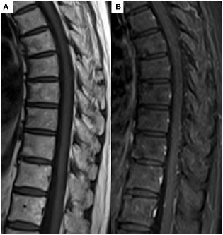
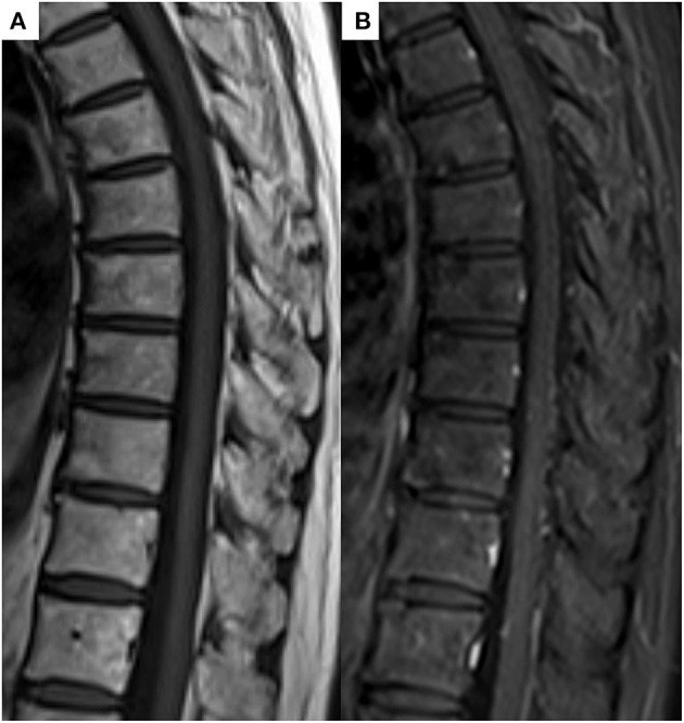
Task. Enhance the resolution of this Magnetic Resonance Imaging image using super-resolution algorithms for improved anatomical detail
Expected output. Higher-resolution MRI with preserved anatomical detail.
Selection note: Low-to-high-resolution sagittal MRI pair.
S10 — style-transfer · Pathology
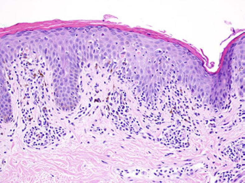
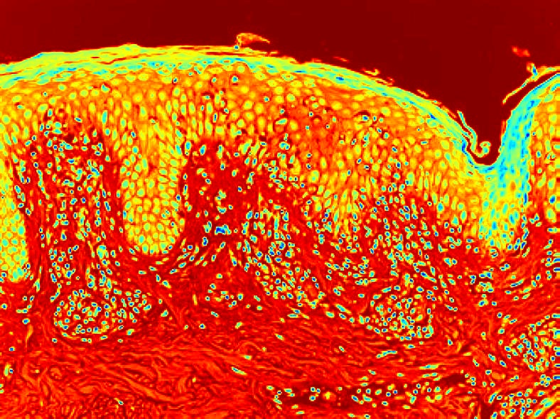
Task. Apply advanced style transfer to this Pathology image while preserving anatomical accuracy and diagnostic quality
Expected output. Thermal-colormap pathology image preserving tissue morphology.
Selection note: Pathology style-transfer pair using a thermal color mapping.
MultimodalGeneration
S11 — 2d-to-3d-reconstruction · CT
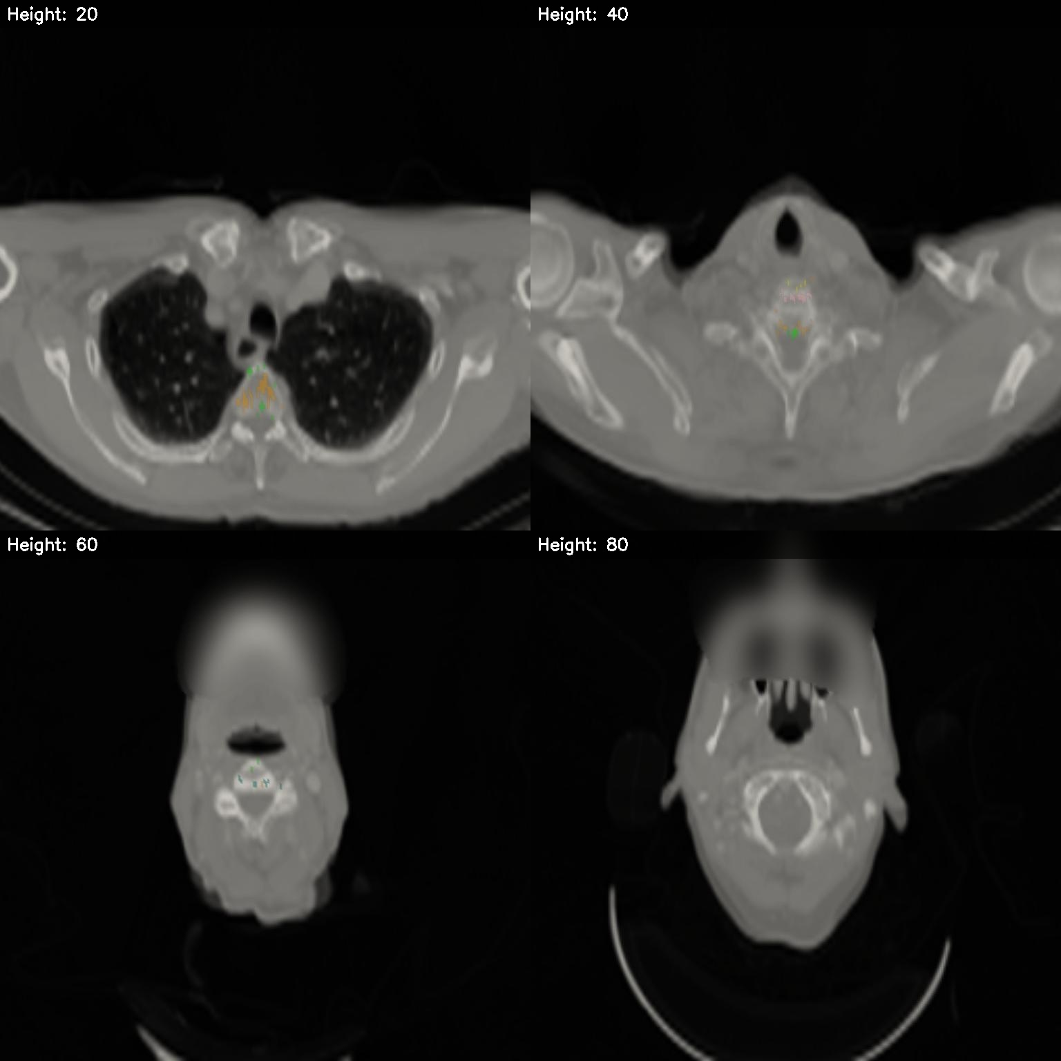
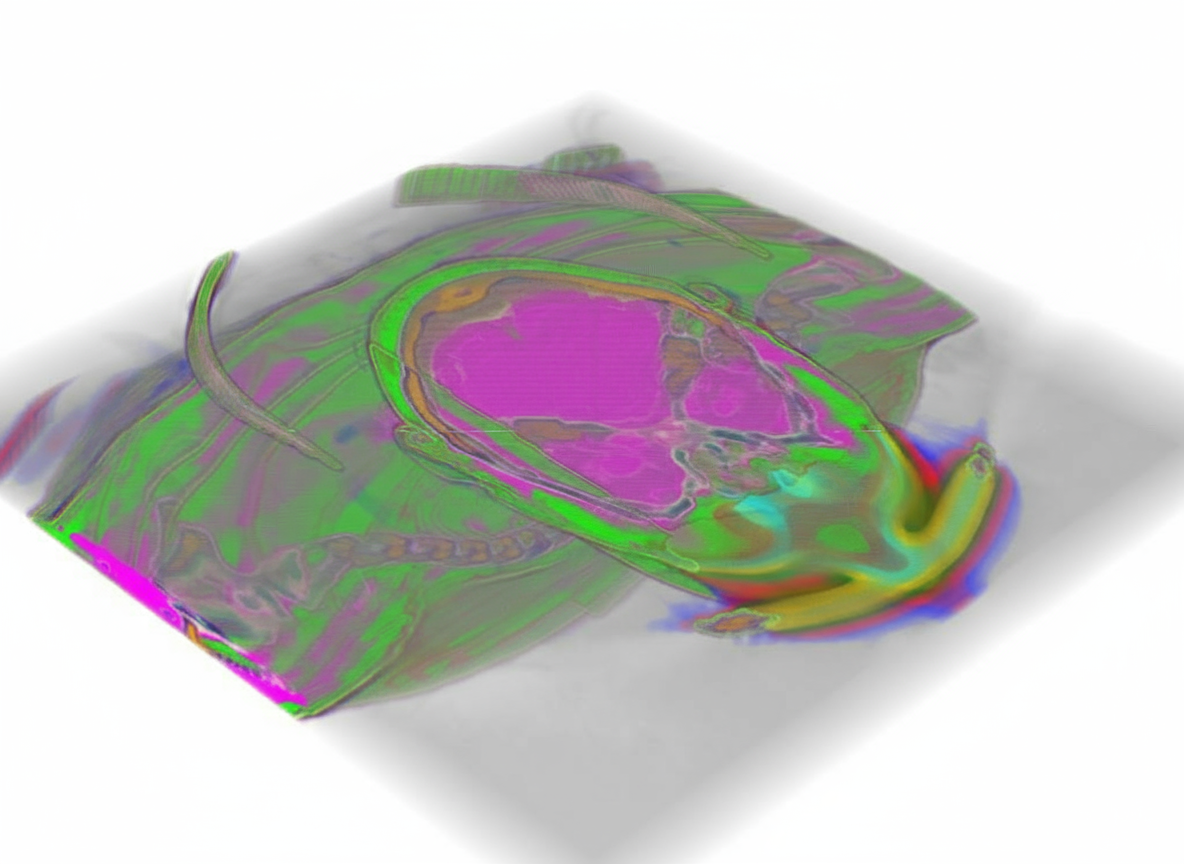
Task. Generate 3D reconstruction from this low attenuation CT slice
Expected output. Color-coded 3D reconstruction of the neck and upper thorax.
Selection note: Multi-slice CT grid to a color-coded 3D reconstruction.
S12 — 3d-to-2d-projection · CT
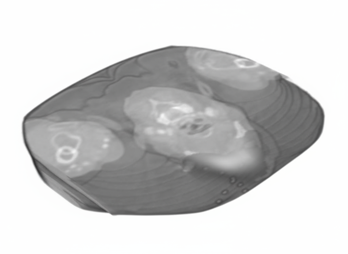
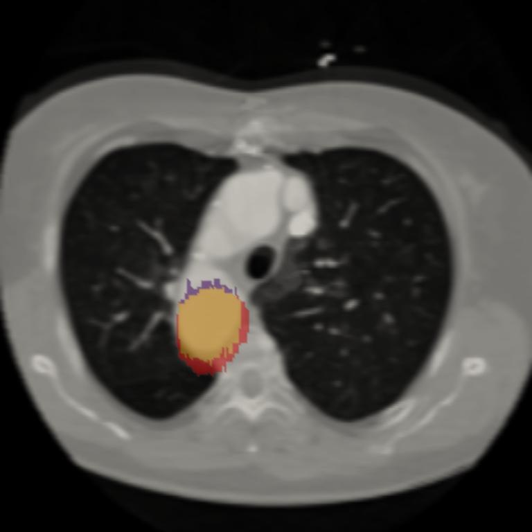
Task. Create axial_cross_section at 100% position in this 3D CT scan
Expected output. Axial chest CT slice with a highlighted right-lung nodule.
Selection note: 3D CT representation to an axial chest slice with a localized nodule.
S14 — instruction-editing · Ultrasound
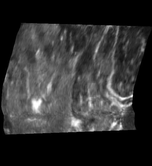
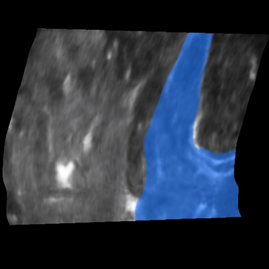
Task. Apply enhancement processing to the jugular_vein using AI enhancement in this hypoechoic Ultrasound longitudinal image
Expected output. Longitudinal thyroid ultrasound with the internal jugular vein segmented in blue.
Selection note: Ultrasound instruction-edit pair with an internal-jugular-vein segmentation overlay.
S15 — organic-reconstruction · Ultrasound
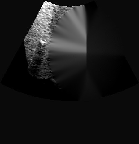
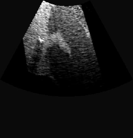
Task. Reconstruct the extensive liver in this speckled Ultrasound image
Expected output. Liver ultrasound reconstructed from a masked region.
Selection note: Masked liver ultrasound reconstructed through an inpainting-style counterfactual edit.
S16 — organic-removal · Other / unspecified
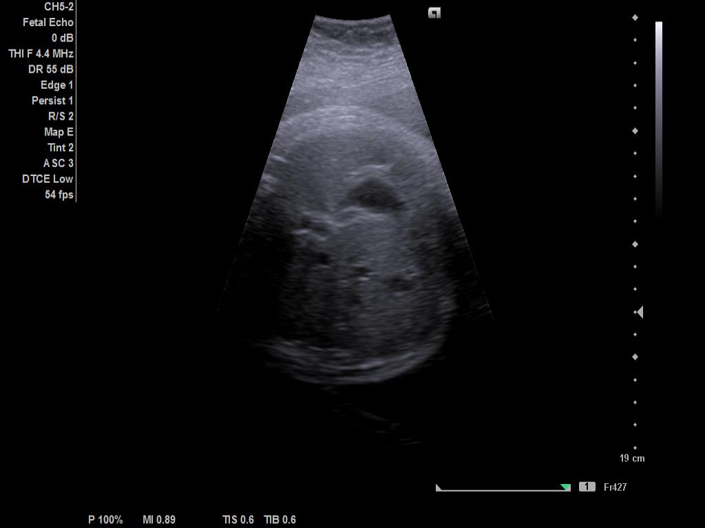
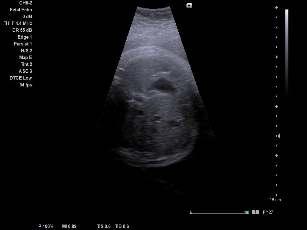
Task. Remove the artery from this medical image using inpainting technique
Expected output. Artery-targeted inpainting counterfactual with surrounding anatomy retained.
Selection note: Fine-grained artery inpainting counterfactual. This is a synthetic editing stress test, not a clinical preprocessing recommendation.
VQA
S17 — multiple-choice · Pathology
 —
—
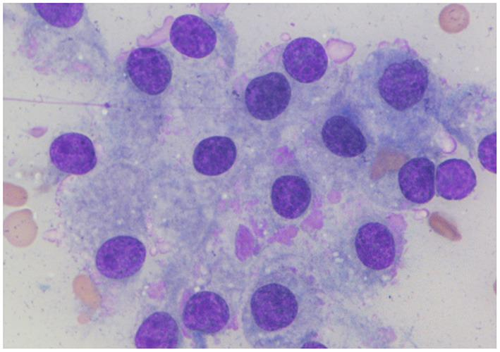
Task. Question: Which organ is this image most likely from? Options: A. Thymus B. Thyroid C. Lymph Node D. Salivary Gland
Expected output. Thyroid.
Selection note: Histopathology multiple-choice item identifying a thyroid tissue structure.
S18 — multiple-choice · Pathology
 —
—
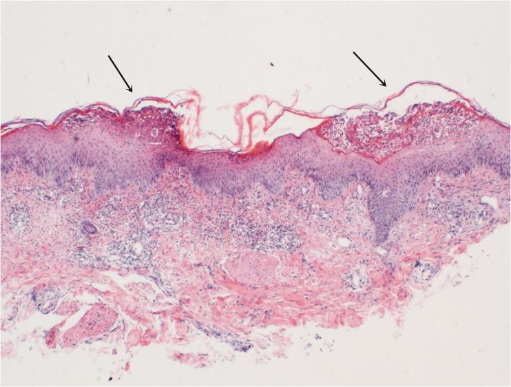
Task. Question: What lesion is most clearly visible in this image? Options: A. Atrophic Rete Ridges B. Hyperkeratosis C. Flat Rete Ridges D. Elongated Rete Ridges E.…
Expected output. Acanthosis.
Selection note: Pathology multiple-choice item with a visible dermatopathology finding.
S19 — blank-filling · Clinical Photography
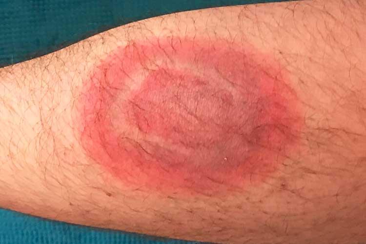 —
—
Task. The pathology shown in this image is __.
Expected output. Erythema migrans.
Selection note: Non-identifying dermatology photograph with a concise pathology answer.
S20 — blank-filling · Clinical Photography
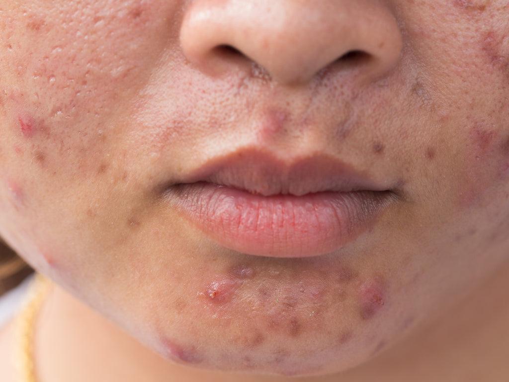 —
—
Task. The pathology is more consistent with __.
Expected output. Acne rosacea.
Selection note: Non-identifying facial-skin crop used for a dermatology blank-filling item.
S21 — question-answering · X-ray
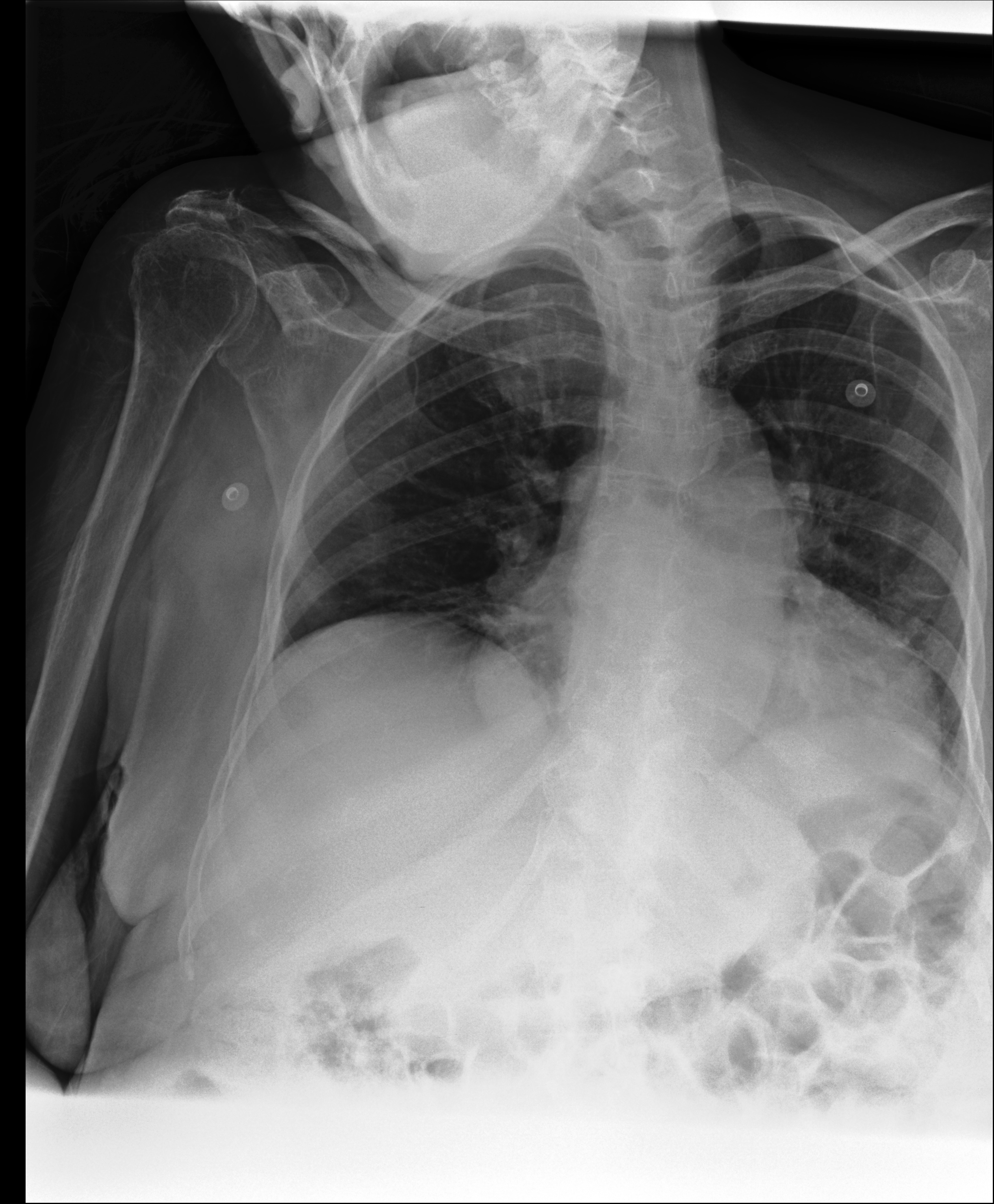 —
—
Task. What imaging modality is used in this image?
Expected output. X-ray.
Selection note: Chest radiograph question asking for the imaging modality.
S22 — question-answering · X-ray
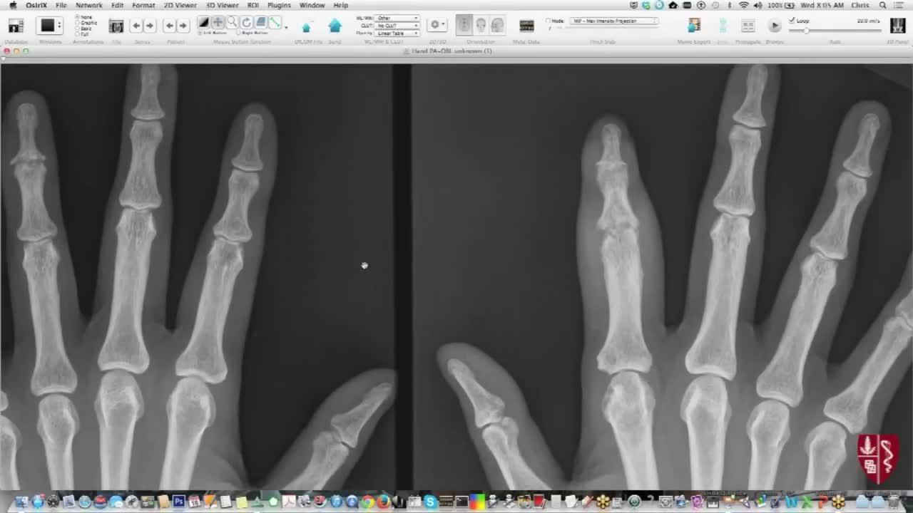
 —
—
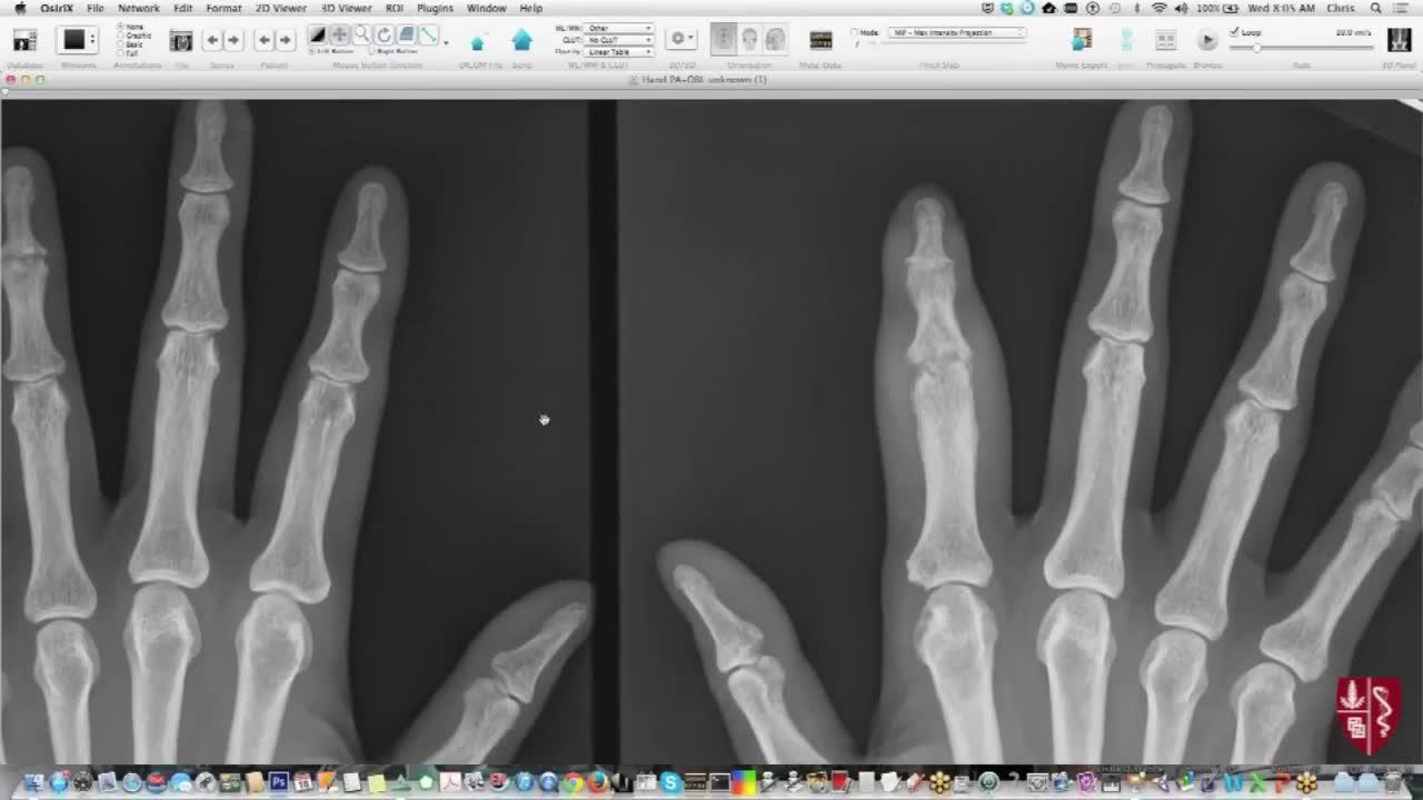
Task. Based on the radiographic findings in the first and second images, which diagnosis is most consistent with the asymmetric involvement of the distal interphalan…
Expected output. Asymmetric distal interphalangeal joint involvement.
Selection note: Multi-view hand radiograph question grounded in distal interphalangeal joints.
S23 — report-generation · CT
 —
—
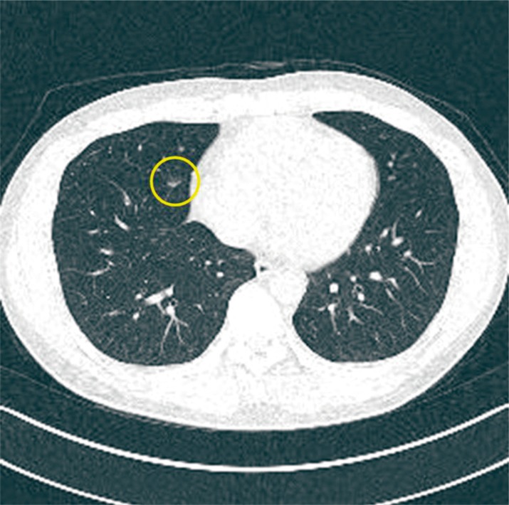
Task. Generate a clinical report for this CT scan.
Expected output. Chest CT report describing small nodules and mediastinal structures.
Selection note: Chest CT report-generation example describing pulmonary nodules.
S24 — report-generation · CT
 —
—
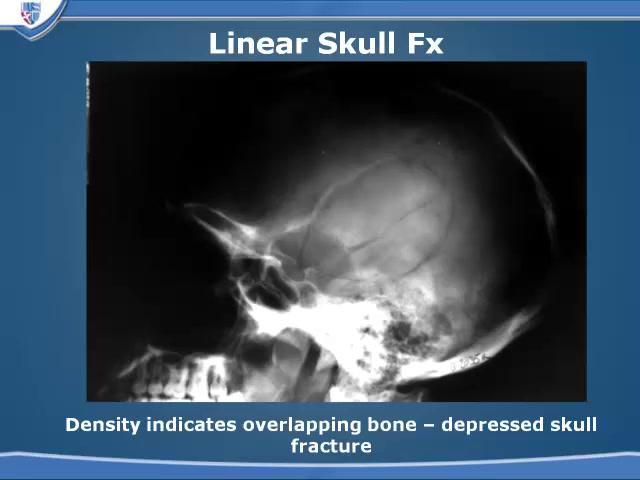
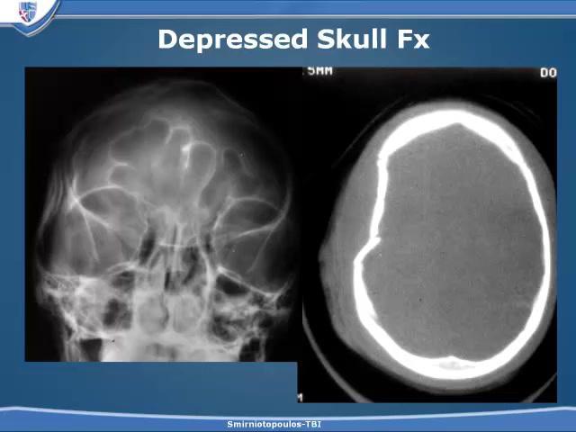
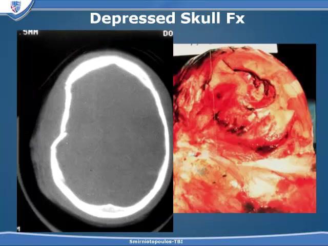
Task. Generate a structured radiology report for the following case: Based on the visual findings across the first, second, and third images, which of the following …
Expected output. Structured report for head trauma and neurological symptoms.
Selection note: Structured radiology-report example using a multi-image skull case.
ImageEditing
S25 — anatomical-annotation · Ultrasound
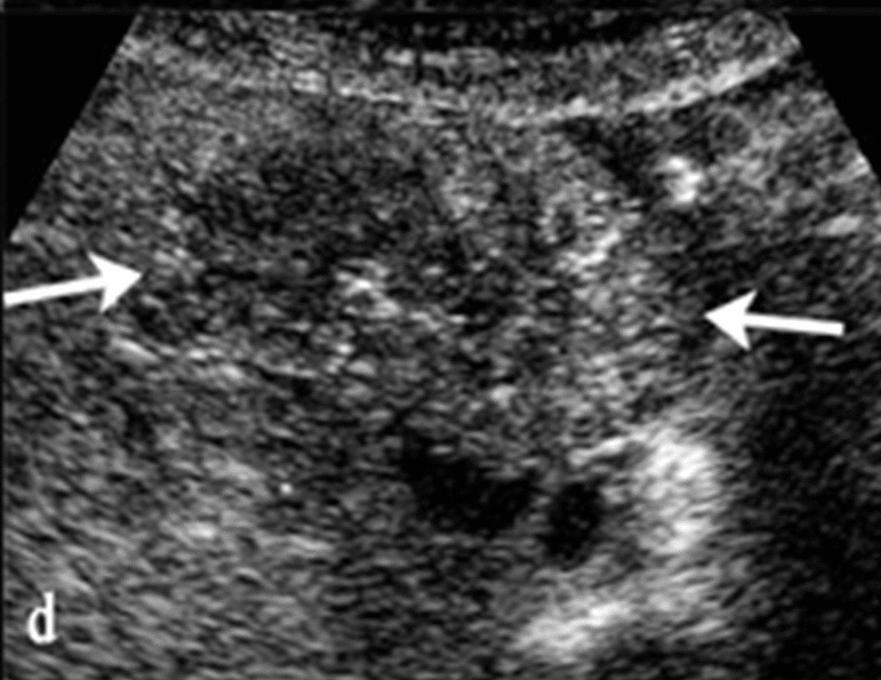
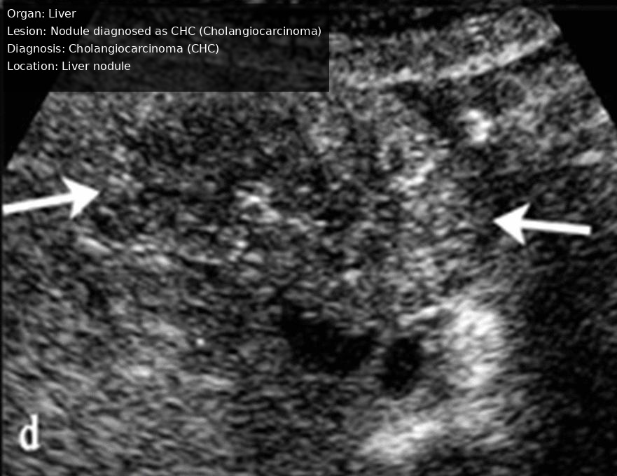
Task. Annotate this Ultrasound image of Liver with detailed labels for the Nodule diagnosed as CHC (Cholangiocarcinoma), including anatomical landmarks and diagnosti…
Expected output. Annotated liver ultrasound with a nodule label.
Selection note: Ultrasound annotation pair for a liver nodule.
S26 — anatomical-annotation · Ultrasound
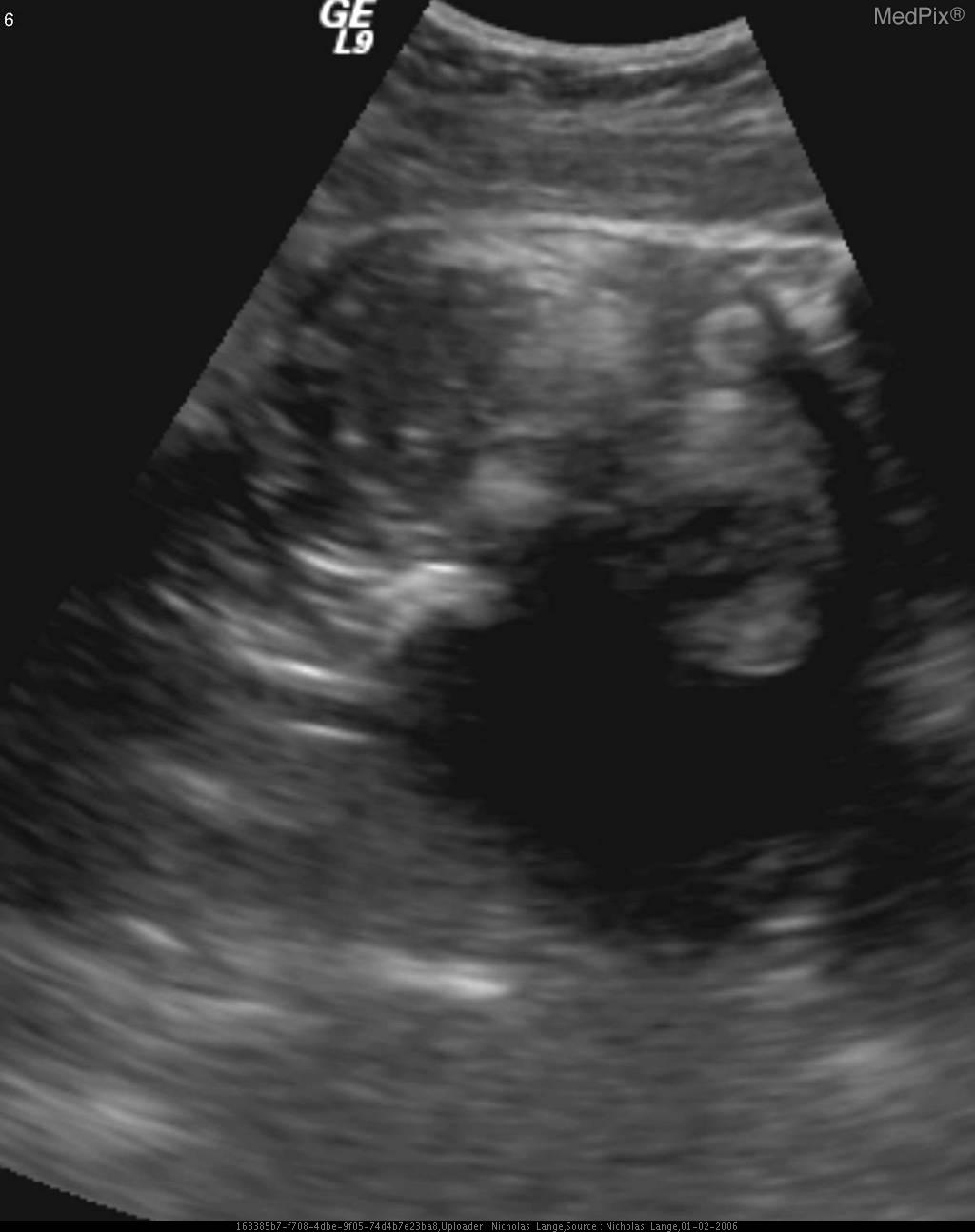
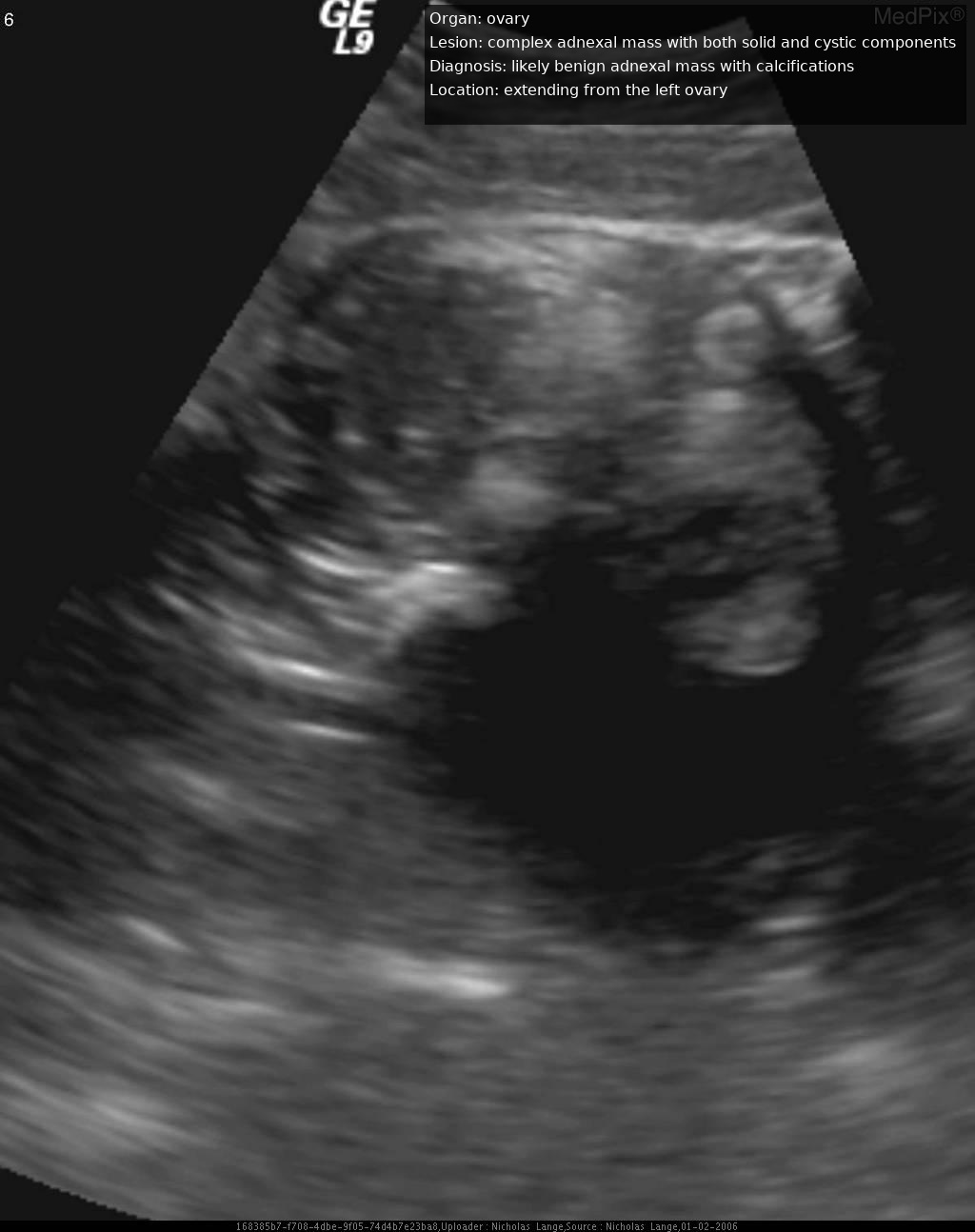
Task. Overlay clinical annotations on this Ultrasound scan
Expected output. Annotated ultrasound image with a lesion-focused label.
Selection note: Ultrasound annotation pair for a complex adnexal lesion.
S27 — artifact-removal · X-ray
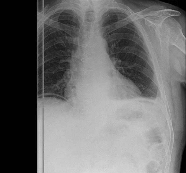
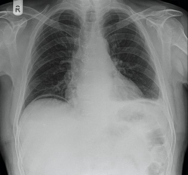
Task. Clean up this X-Ray scan
Expected output. Post-artifact-removal PA chest radiograph.
Selection note: Validated chest-radiograph artifact-removal example with truncation lines removed.
S28 — artifact-removal · X-ray
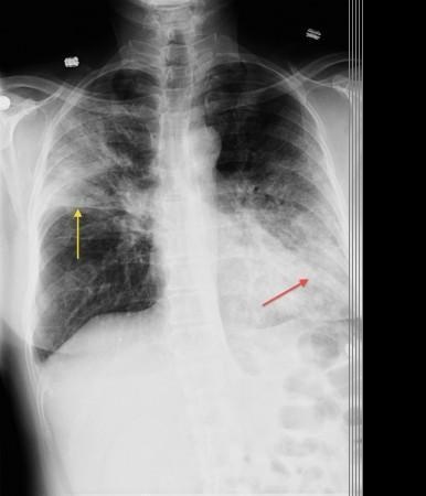
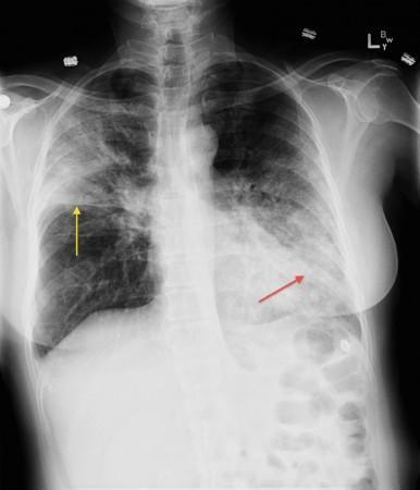
Task. Eliminate distortions from this X-Ray scan
Expected output. Chest X-ray with the left breast shadow digitally reduced.
Selection note: Validated X-ray artifact-removal example showing improved lung-field visibility.
S29 — contrast-enhancement · X-ray
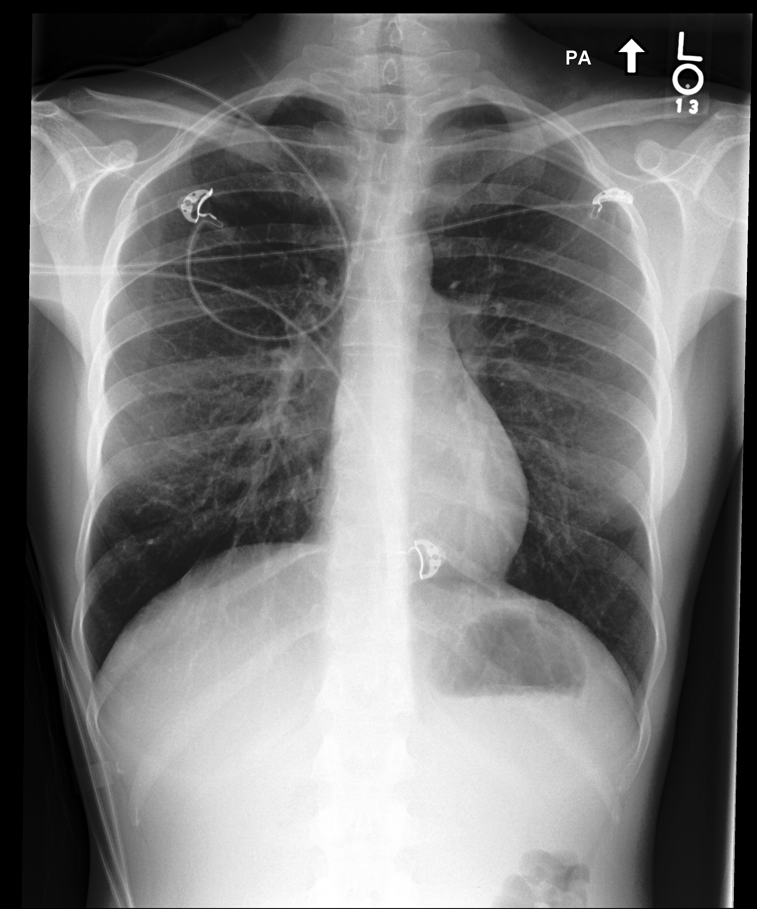
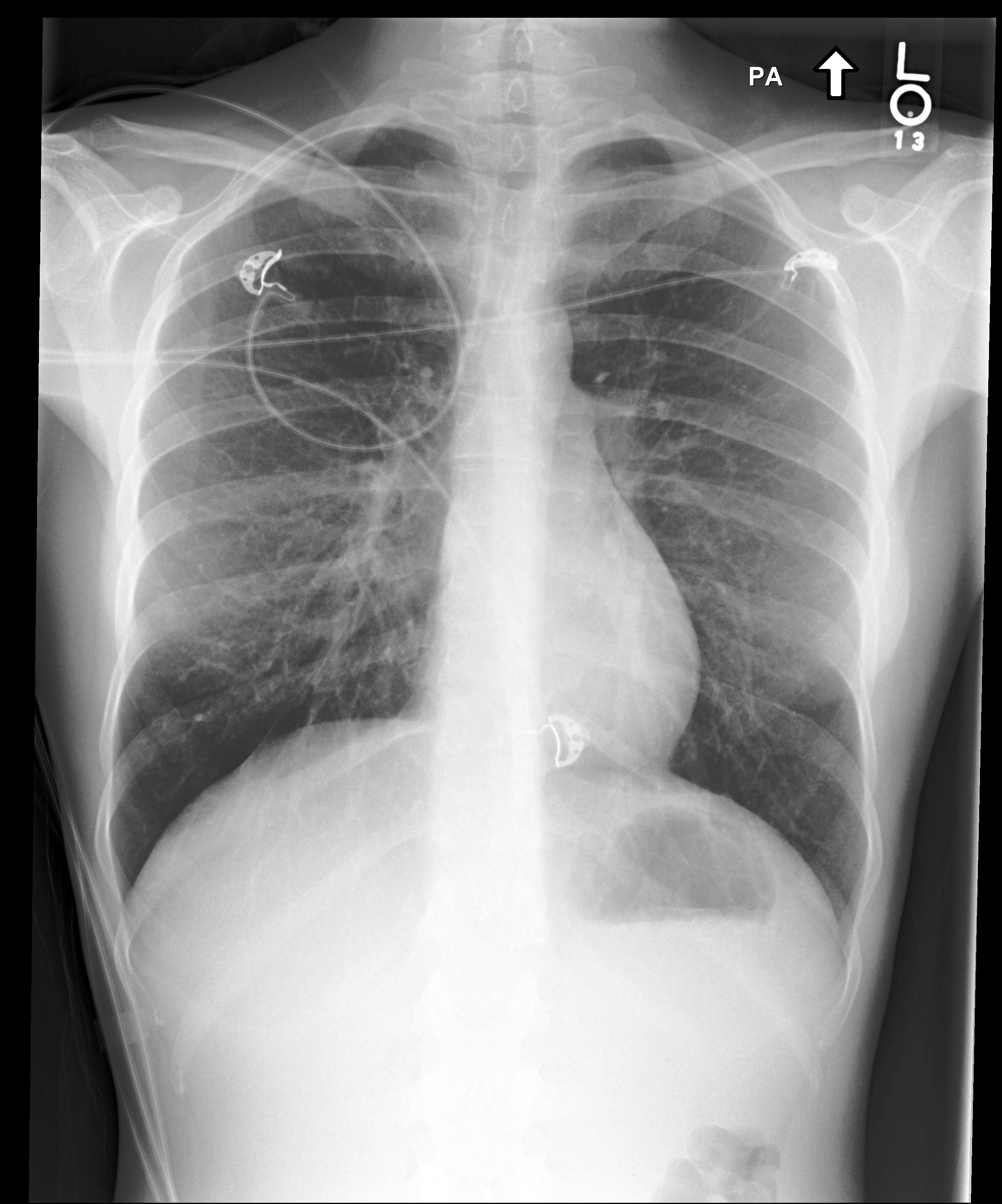
Task. Improve the visual quality of this Radiograph scan through advanced contrast enhancement while preserving diagnostic information
Expected output. Contrast-enhanced X-ray with improved anatomical visibility.
Selection note: X-ray contrast-enhancement pair with preserved anatomical structures.
S30 — contrast-enhancement · Clinical Photography
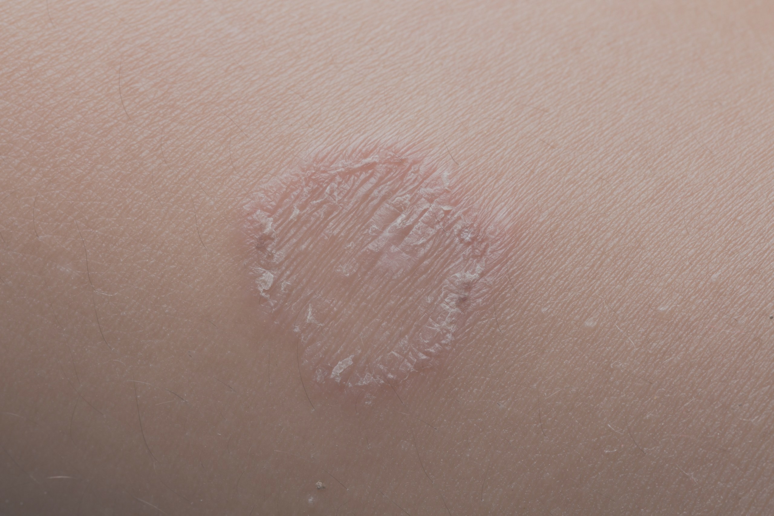
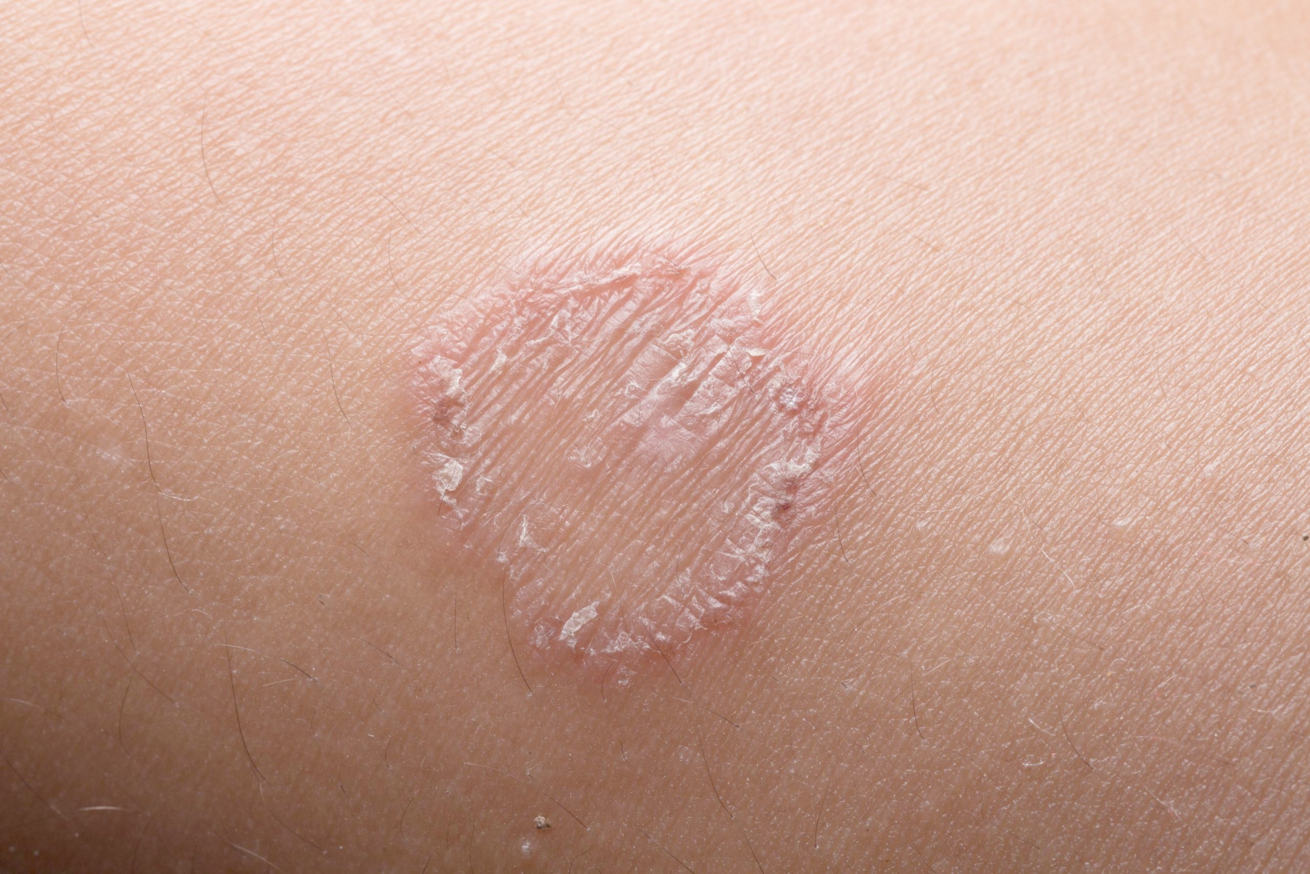
Task. Enhance the contrast of this Clinical Photo image using advanced image processing algorithms for improved diagnostic visibility
Expected output. Skin lesion with darker, more diagnostic contrast.
Selection note: Non-identifying dermatology contrast-enhancement pair.
S31 — noise-reconstruction · CT
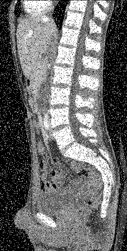
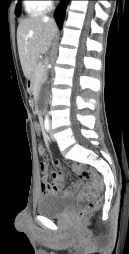
Task. Apply sophisticated noise reduction to this CT scan while preserving fine anatomical details and diagnostic features
Expected output. Denoised CT sagittal view with preserved vertebral anatomy.
Selection note: Salt-and-pepper-noise reduction on a CT sagittal view.
S32 — noise-reconstruction · CT
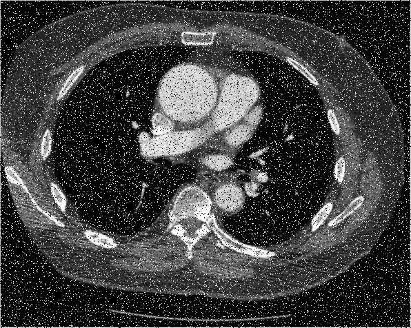
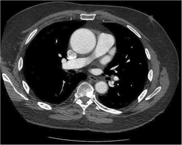
Task. Clean up this noisy CT image
Expected output. Cleaner chest CT with preserved cardiopulmonary structures.
Selection note: Chest CT cleanup example with visible noise reduction.
S33 — resolution-editing · MRI
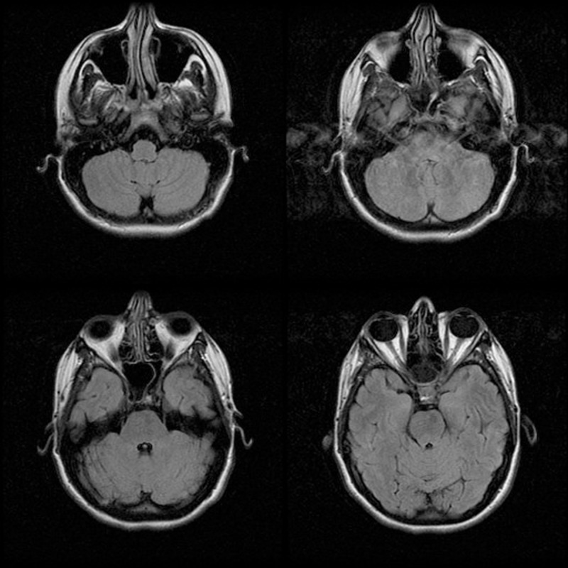
Task. Improve the spatial resolution of this Magnetic_Resonance_Imaging image using deep learning-based super-resolution methods
Expected output. Higher-resolution multi-view MRI.
Selection note: Multi-view brain MRI resolution-enhancement pair.
S34 — resolution-editing · MRI
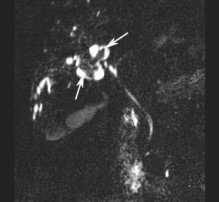
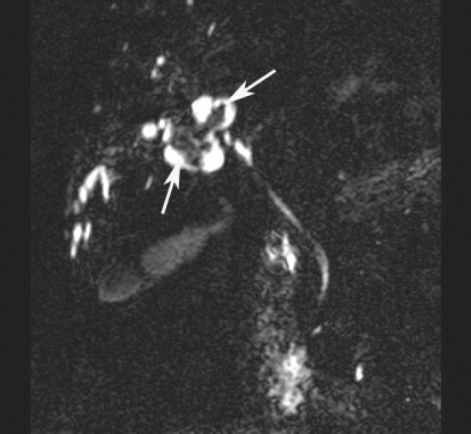
Task. Enhance the resolution of this low-quality Magnetic Resonance Imaging image
Expected output. MRI with enhanced spatial detail.
Selection note: Focused MRI resolution-enhancement pair with a visible anatomical target.
S35 — style-transfer · Pathology
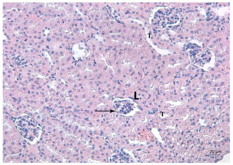
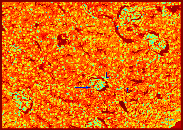
Task. Transform this Pathology using thermal colormap visualization to enhance tissue contrast and signal intensity distribution. Technical Implementation: Apply the…
Expected output. Thermal-colormap pathology visualization.
Selection note: Pathology thermal-colormap style-transfer pair with strong visual contrast.
S36 — style-transfer · Pathology
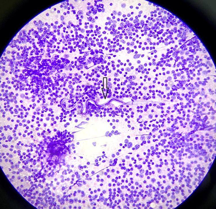
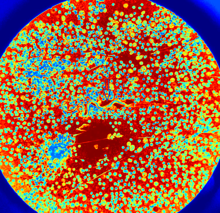
Task. Transform this Pathology using thermal colormap to enhance tissue contrast. Technical: Apply thermal mapping (blue-cold to red-hot) with histogram equalization…
Expected output. Thermal-mapped pathology image.
Selection note: Pathology thermal-mapping pair with preserved tissue layout.
MultimodalGeneration
S37 — 2d-to-3d-reconstruction · CT
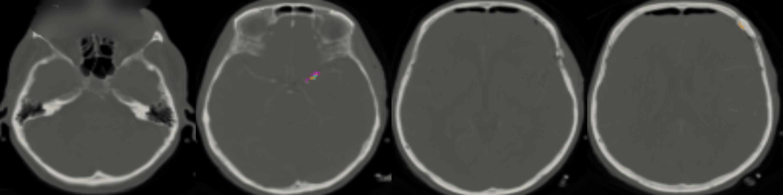
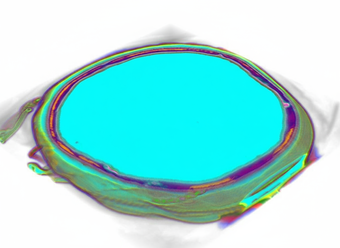
Task. Reconstruct 3D volume from this 2D CT image
Expected output. Color-coded 3D brain-parenchyma reconstruction.
Selection note: CT slices converted into a color-coded 3D segmentation view.
S38 — 2d-to-3d-reconstruction · CT
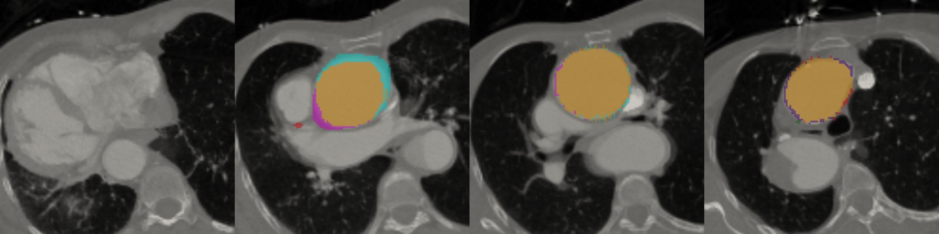
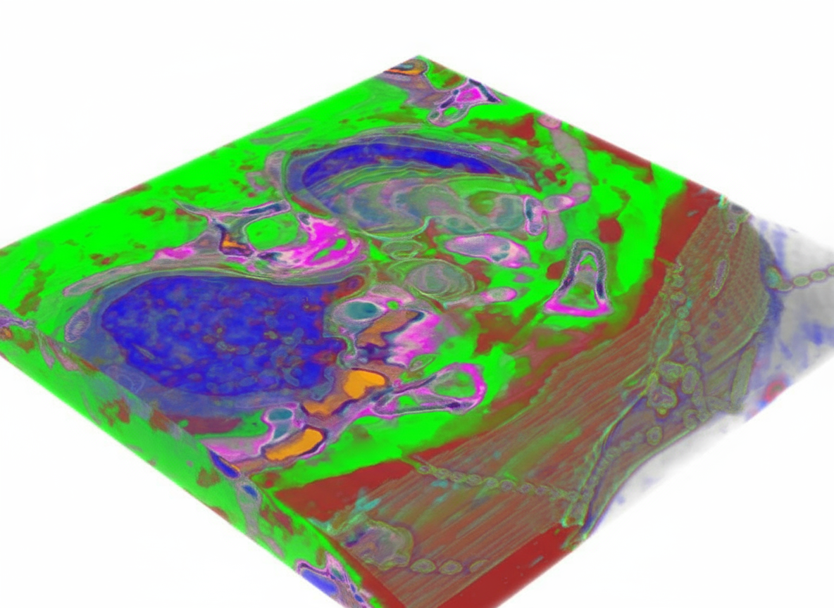
Task. Create volumetric model from this 2D CT scan
Expected output. Color-coded 3D thoracic reconstruction.
Selection note: Multi-slice CT grid converted into a color-coded 3D thoracic model.
S39 — 3d-to-2d-projection · CT
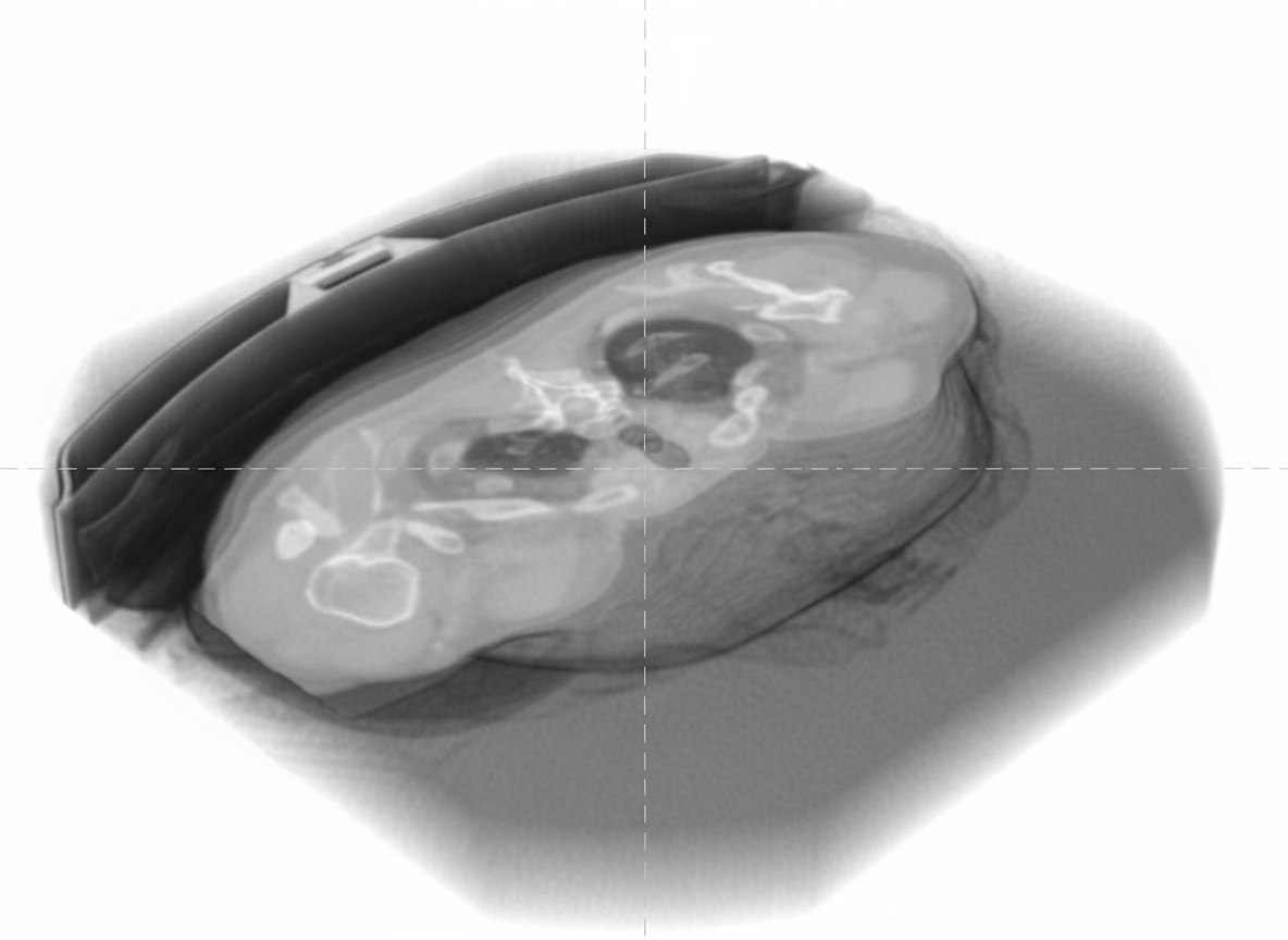
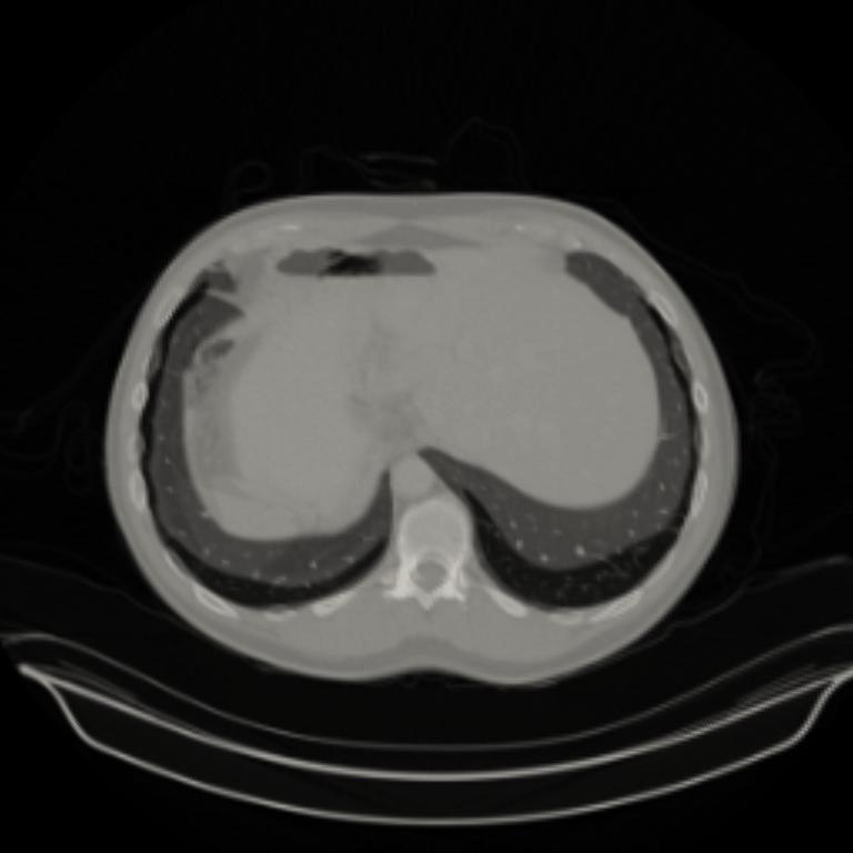
Task. Create axial_cross_section at 75% position in this 3D CT scan
Expected output. Axial upper-abdominal CT projection.
Selection note: Grayscale axial projection from a 3D CT representation.
S40 — 3d-to-2d-projection · CT
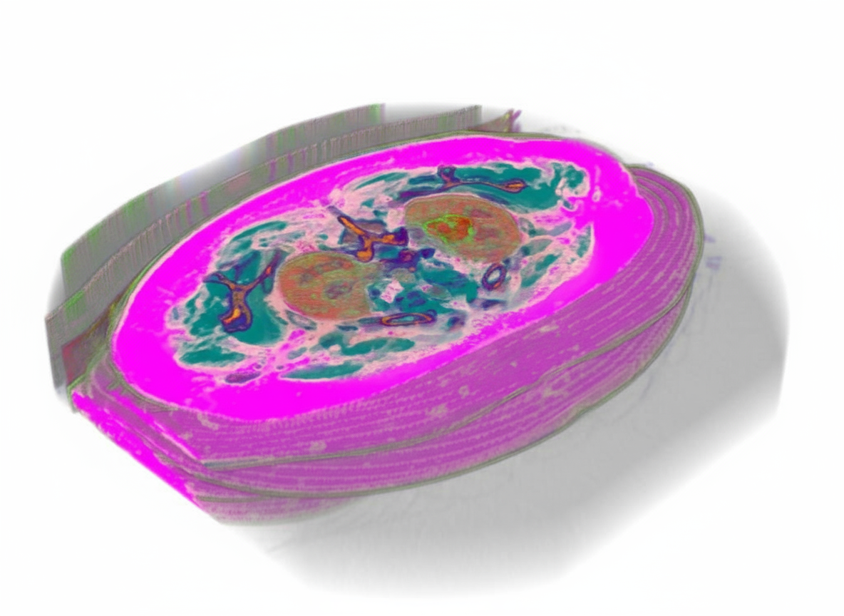
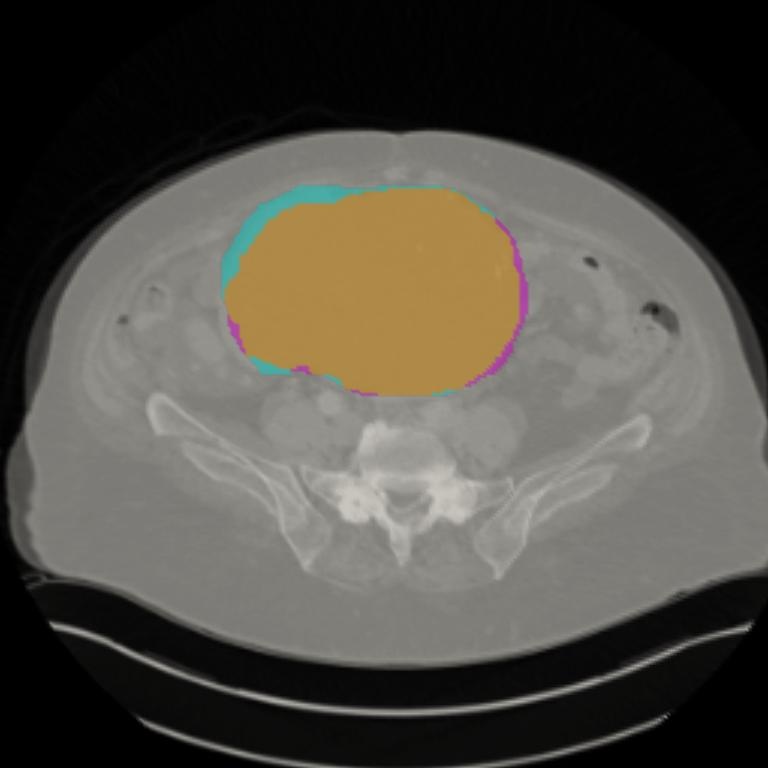
Task. Create axial_cross_section at 50% position in this 3D CT scan
Expected output. Axial CT slice with a segmented bladder.
Selection note: Color-coded 3D CT representation projected to a labeled axial slice.
S43 — instruction-editing · Ultrasound
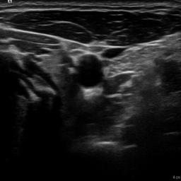
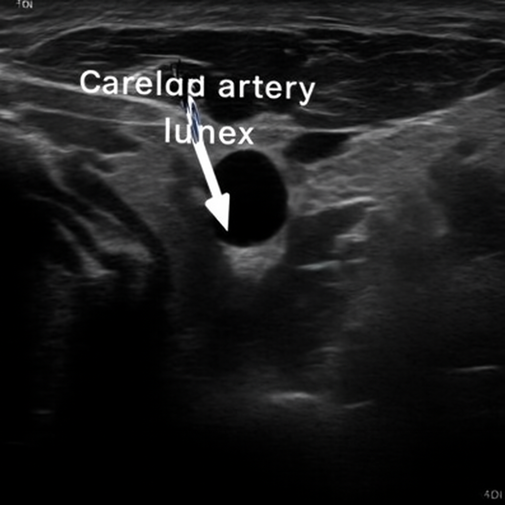
Task. Process the carotid artery lumen with AI enhancement in this heterogeneous Ultrasound longitudinal view
Expected output. Longitudinal carotid ultrasound with the lumen labeled.
Selection note: Carotid-lumen instruction-edit pair with a visible label overlay.
S44 — instruction-editing · Ultrasound
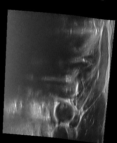
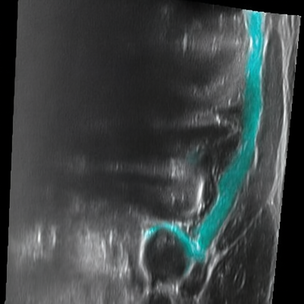
Task. Apply AI-based enhancement enhancement to the carotid_artery in this heterogeneous Ultrasound longitudinal image
Expected output. Longitudinal neck ultrasound with the carotid artery segmented.
Selection note: Common-carotid instruction-edit pair with a blue segmentation overlay.
S45 — organic-reconstruction · Ultrasound
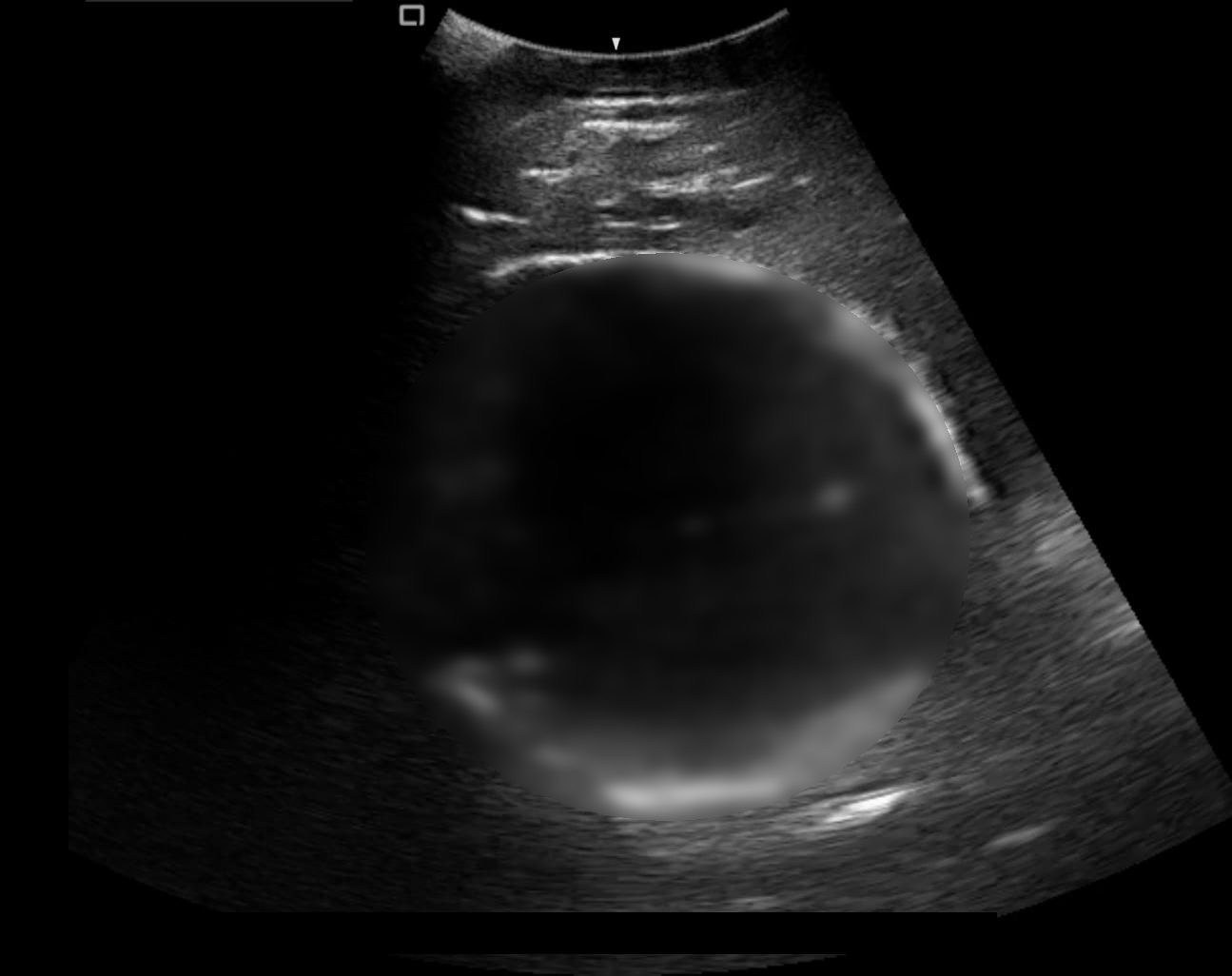
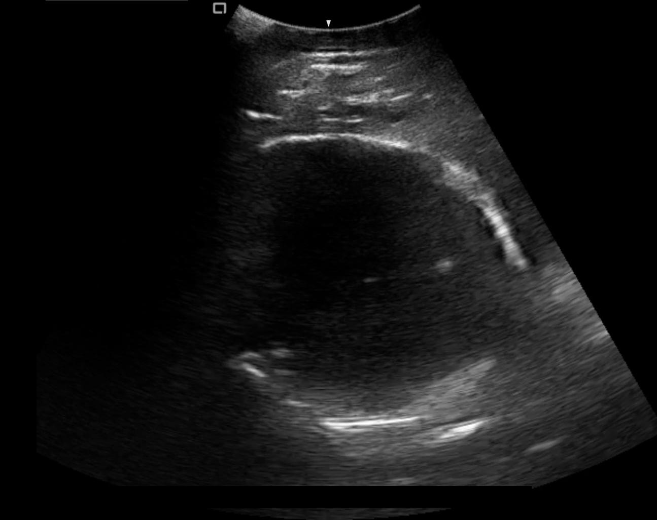
Task. Reconstruct the large-scale fetal head in this homogeneous Ultrasound image
Expected output. Fetal-head ultrasound reconstructed from a masked region.
Selection note: Fetal-head ultrasound reconstructed from an obscured region.
S46 — organic-reconstruction · Ultrasound
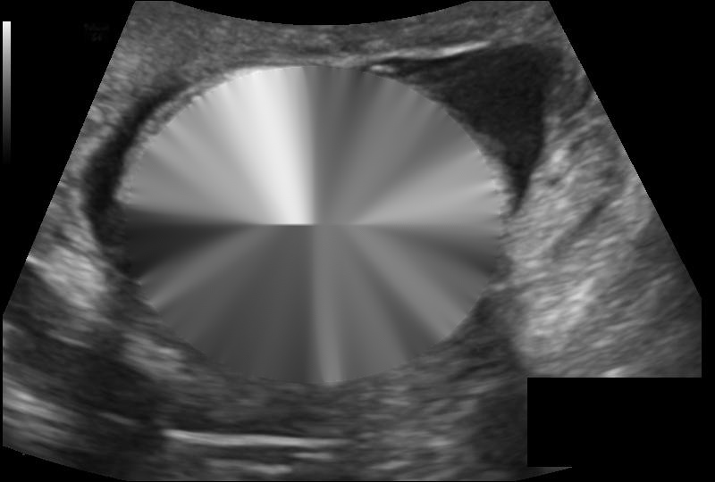
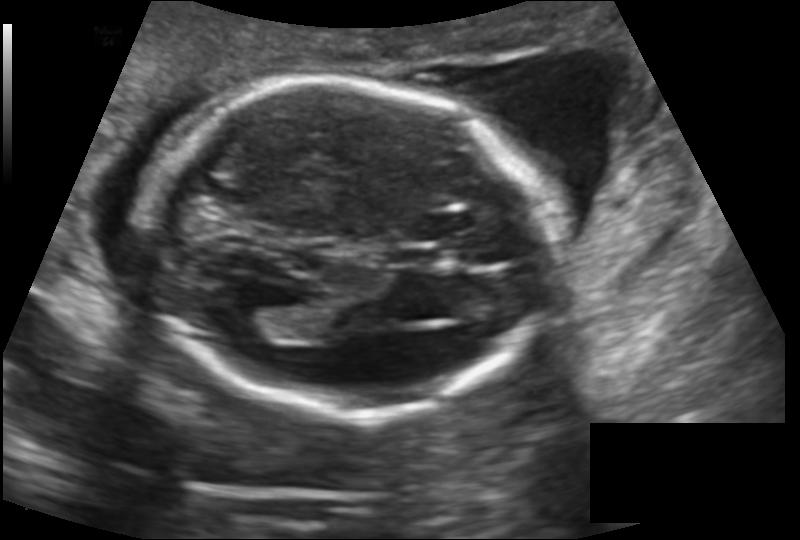
Task. Reconstruct the extensive head_circumference in this anechoic Ultrasound image
Expected output. Reconstructed fetal-head ultrasound with clear cranial structures.
Selection note: Fetal-head ultrasound reconstruction with the cranial boundary restored.
S47 — organic-removal · Other / unspecified
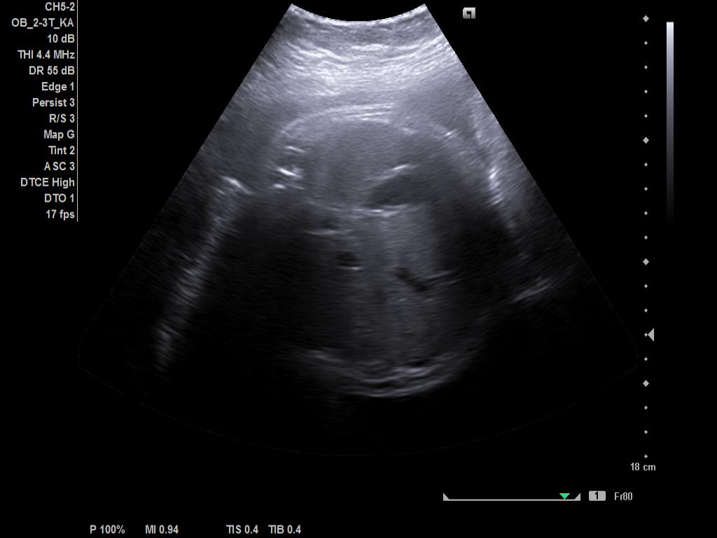
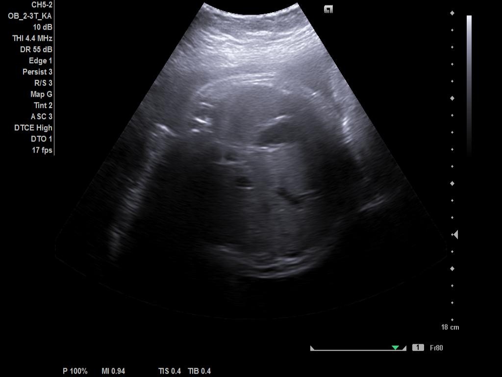
Task. Remove the artery from this medical image using inpainting technique
Expected output. Artery-targeted inpainting counterfactual.
Selection note: Artery-targeted inpainting counterfactual retained for task-level inspection.
S48 — organic-removal · Other / unspecified
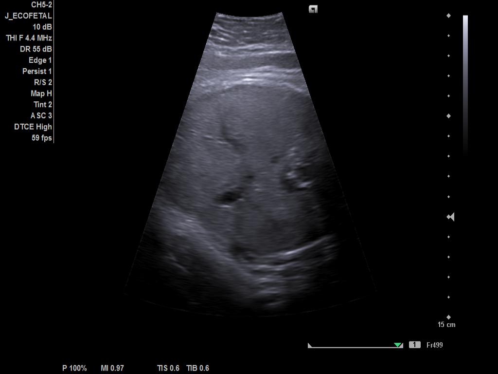
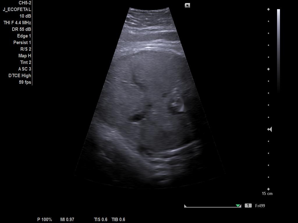
Task. Remove the artery from this medical image using inpainting technique
Expected output. Artery-removal inpainting counterfactual.
Selection note: Artery-removal inpainting counterfactual with native-resolution ultrasound context.
Review-only conditions
The package contains no author names, e-mail addresses, API credentials, account identifiers, absolute workstation paths, or source-code files. Source-dataset labels and non-identifying record metadata are retained only to enable audit of task provenance. Any downstream reuse remains subject to the original source-data terms and approvals.
For reviewer use, upload this ZIP with the file designation Dataset. TMI permits datasets as Supporting Documents, while it does not permit a supplementary manuscript-like document that extends the main paper with prose or figures. The 40 MB TMI limit applies to the manuscript PDF; this package is deliberately kept below 39 MiB as a conservative portal-safe target.